Imaging
Diagnosis in Sight
From Detection to Progression
Imaging plays a critical role in both early diagnosis of GA and in the monitoring of disease progression.
Diagnostic Hallmarks of AMD
Early
Multiple small (<63 µm) and few intermediate
(63-124 µm) drusen, or RPE abnormalities1
Intermediate
Extensive intermediate drusen (63-124 µm) or more than 1 large druse (≥125 µm).1 May also be accompanied by degenerative changes in the choriocapillaris, RPE, and photoreceptors1-3
Advanced
Progressive atrophy of choriocapillaris, RPE, and photoreceptors.2,3 As well as new and growing lesions3
Optical Coherence Tomography (OCT) | Color Fundus Photography (CFP) | Fundus Autofluorescence (FAF) |
Intermediate AMD | Intermediate AMD | Intermediate AMD |
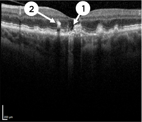 Image courtesy of Dr. Arshad Khanani |  Image courtesy of Dr. Arshad Khanani | 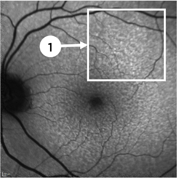 Image courtesy of Dr. Arshad Khanani |
Advanced AMD (Geographic Atrophy) | Advanced AMD (Geographic Atrophy) | Advanced AMD (Geographic Atrophy) |
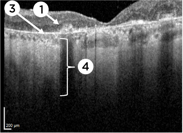 Image courtesy of Dr. Arshad Khanani | 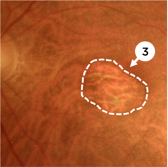 Image courtesy of Dr. Mohammad Rafieetary | 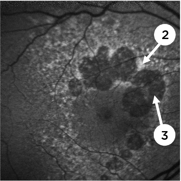 Image courtesy of Dr. David Lally |
Key Features1,4
Images are from separate patients. | Key Features4-6
Images are from separate patients. | Key Features1,7
Images are from separate patients. |
Optical Coherence Tomography (OCT)
Intermediate AMD
Image courtesy of Dr. Arshad Khanani
Advanced AMD (Geographic Atrophy)
Image courtesy of Dr. Arshad Khanani
Key Features1,4
- (1) Degeneration of overlying photoreceptors increases reflectivity below Bruch’s membrane
- (2) Hyperreflective foci corresponds to attenuation or disruption of the RPE
- (3) Atrophy of the RPE
- (4) Area of choroidal hypertransmission
Images are from separate patients.
Color Fundus Photography (CFP)
Intermediate AMD
Image courtesy of Dr. Arshad Khanani
Advanced AMD (Geographic Atrophy)
Image courtesy of Dr. Mohammad Rafieetary
Key Features4-6
- (1) Drusen and (2) fundus abnormalities are identifiable as hyperpigmented areas
- (3) GA lesion borders are sharply demarcated with increased choroidal vessel visibility (dashed circle)
Images are from separate patients.
Fundus Autofluorescence (FAF)
Intermediate AMD
Image courtesy of Dr. Arshad Khanani
Advanced AMD (Geographic Atrophy)
Image courtesy of Dr. David Lally
Key Features1,7
- (1) Reticular pseudodrusen appearing as multiple, clustered, regularly networked, round areas of low-contrast hypo-autofluorescence may be prognostic of advancing AMD
- (2) Areas of hypoautofluorescence with sharply demarcated borders indicate atrophic lesions
- (3) Abnormal patterns of hyperautofluorescence surrounding atrophic lesions
Images are from separate patients.
Watch and Discover
Find out more about imaging GA, such as key diagnostic and monitoring hallmarks, from your colleagues in this video. Or by downloading the Imaging Guide for Early Detection and Monitoring, linked below.
Imaging Guide for Early Detection and Monitoring

Importance of Imaging
Importance of Imaging
References
- Fleckenstein M, Mitchell P, Freund KB, et al. The progression of geographic atrophy secondary to age-related macular degeneration.Ophthalmology. 2018;125(3):369-390.
- Holz FG, Schmitz-Valckenberg S, Fleckenstein M. Recent developments in the treatment of age-related macular degeneration. J Clin Invest. 2014;124(4):1430-1438.
- Boyer DS, Schmidt-Erfurth U, van Lookeren Campagne M, Henry EC, Brittain C. The pathophysiology of geographic atrophy secondary to age-related macular degeneration and the complement pathway as a therapeutic target.Retina. 2017;37(5):819-835.
- Sadda SR, Guymer R, Holz FG, et al. Consensus definition for atrophy associated with age-related macular degeneration on OCT. Ophthalmology. 2018;125(4):537-548.
- Sadda SR, Chakravarthy U, Birch DG, Staurenghi G, Henry EC, Brittain C. Clinical endpoints for the study of geographic atrophy secondary to age-related macular degeneration. Retina. 2016;36(10):1806-1822.
- Garrity ST, Sarraf D, Freund KB, Sadda SR. Multimodal imaging of nonneovascular age-related macular degeneration. Invest Ophthalmol Vis Sci. 2018;59(4):AMD48-AMD64.
- Yung M, Klufas MA, Sarraf D. Clinical applications of fundus autofluorescence in retinal disease. Int J Retina Vitreous. 2016;2:12.